Should stethoscopes be displayed in museums?

Stanford Healthcare Innovation Team
Feb 3, 2022

Get in touch

Reach out to contribute a post like this.

The medical profession is closely associated with stethoscopes. For example, googling ‘doctor’ results in images of stethoscopes, or doctors wearing stethoscopes. A 2012 study showed that physicians are much more likely to be regarded as ‘trustworthy’ if they have a stethoscope with them [1]. However, with rapid technological advancements and the miniaturization of sensors and electronics, stethoscopes might find their way into museums in the near future.The Evolution of Stethoscopes

The concept of a stethoscope was first realized in 1816 by René Théophile Hyacinthe Laënnec. He noticed children amplified sounds by playing with hollow sticks, and this observation gave him the idea of using a tube to auscultate patients. His first stethoscope was a simple paper tube used to listen to heartbeats, but he experimented with different materials and eventually settled on a wooden tube with a plug on the end. Unfortunately, this tube only had listening access for one ear.

Leftmost, a diagram of Laënnec’s stethoscope model is shown. In the middle, Laënnec’s stethoscope is shown. Rightmost, Laënnec is shown using his stethoscope to auscultate a patient. Image credit: National Center for Biotechnology Information

By 1843, stethoscopes were upgraded and doctors could now listen with both ears. By 1945, the modern design of the stethoscope was finalized using thin tubing, rubber earpieces, and more features. By 1956, stethoscopes were being made with modern materials such as PVC or latex. By 2019, noise cancellation technology, and microphones had been successfully adapted into stethoscopes [2]. With the new technology also came a price increase. A stethoscope’s cost on average is $100, but there is a range of prices. Disposable ones are as little as $5, good stethoscopes are $75, and high-end gadgetry can push the cost to $300 [34].

From the 1970s onward, electronics have revolutionized medicine, from being able to request medications remotely to genetic sequencing. Even the stethoscope has received upgrades. Recently, Eko [5] designed an add-on to a stethoscope that can detect abnormal pulmonary and heart conditions, has noise-canceling technology and links to Bluetooth. Other possible innovations (which are not widely used) include on-the-spot AI analysis, connecting ECG probes to a stethoscope, and replacing the stethoscope with a listening device and wireless earbuds. Companies, such as Thinklabs [6] are already working on such alterations. These innovations to the stethoscope would allow doctors to obtain additional diagnostic information. Adding ECG probes can also alert to abnormal electrical activity in the heart, and allow better diagnoses for arrhythmia. An example of mobile ECG technology approved by the FDA (which could be integrated into future stethoscopes) is in the KardiaMobile offered by AliveCor [7].

A cardiology grade stethoscope with Eko Health’s electronic add-on. Image credit: Eko Health

Limitations of Today’s Stethoscopes

The stethoscope works by ‘listening’ to the body sounds when its diaphragm is pressed against the skin. Applying more pressure on the diaphragm would enable the user to hear higher frequencies.[8]. In this case, low-frequency sounds include  Korotkoff sounds, arrhythmias, while high-frequency sounds include airflow through the lungs [9].

Using a stethoscope. Image credit: Littmann

However, stethoscopes are not without inherent limitations. The stethoscope may indicate if the heart or lungs are diseased, but doesn’t provide any further information for a conclusive diagnosis [10]. Stethoscopes also can’t facilitate virtual diagnosis, which is growing especially important due to the ongoing COVID-19 pandemic.  To receive an interpretation from a stethoscope, a doctor has to be physically present and close to the patient, which is a hindrance when doctors are placed in unstable situations or where patients are immobile or in remote locations. Even with the enhancements stated earlier, some of the above limitations would still apply.

Solutions

There are different paths for improvements. One path forward could be to incrementally enhance the stethoscope. Companies such as Eko, Thinklabs, and StethoMe have already commenced such enhancements. These enhancements would slowly transition towards replacing the stethoscope entirely. For such a change to occur, the replacement must have the functionalities of a modern stethoscope, eliminate its limitations, and incorporate additional features.

There have been proposals to replace stethoscopes with handheld ultrasound devices [11], which give images of the internal human body. A stethoscope allows doctors only to hear the sound, while an ultrasound allows doctors to “see the sound” [12]. It poses almost no health risk because it does not use any form of radiation. Instead, it uses high-frequency sound waves between 2-18 megahertz [13]. Although lower frequency sound waves can penetrate farther into the body, they produce blurrier images. Higher frequency sound waves produce good quality images but get absorbed by tissues more easily. Since ultrasounds cannot penetrate through the bone, they are not generally used for brain imaging.  An ultrasound can be used to diagnose conditions in the lungs, liver, gallbladder, spleen, pancreas, kidneys, bladder, uterus, ovaries, eyes, thyroid, and testicles [14]. Doppler ultrasounds can even determine the velocity of blood flow thereby enabling detection of clots, heart disease, and other cardiovascular conditions [15]. Doppler ultrasounds work by measuring the magnitude of ‘red’ or ‘blue’ (Doppler) shift of echoed frequencies to determine the velocity of the target [16]. The figure below demonstrates a Doppler ultrasound showing deep vein thrombosis of the subsartorial vein.

The blue shows blood flow from the deep femoral vein into the common femoral vein, while there is a total absence of blood flow from the subsartorial vein. The red shows blood flow through the femoral artery. Image credit: Wikipedia

The bulky ultrasound machines driven into cramped hospital wards are transitioning to handheld devices whose images can be displayed on cellphones and laptops thanks to advancements and miniaturization of electronics [17]. Handheld ultrasounds can diagnose a larger variety of conditions than stethoscopes can [18]. For example, stethoscopes cannot detect small tumors in the lungs [19], while ultrasounds are able to locate them, as shown in the figure below.

Seeing the sound: subpleural lung mass, sharply demarcated from well-ventilated lung tissue, seen through ultrasound. Image credit: Medical Ultrasonography

Ultrasounds are more sensitive (97%) & specific (98%) in diagnosing heart failure than stethoscopes (46% and 67%, respectively) [20]. Imaging enables better medical interpretation than simply listening to sounds in a patient’s body. In a study, medical students with 18 hours of cardiac ultrasound training and board-certified cardiologists diagnosed 61 patients with clinically significant cardiac disease. The medical students correctly identified 75% of pathologies, compared to just 49% for the cardiologists [21]. Stethoscopes, on the other hand, are useful for quickly diagnosing conditions that produce sound in a patient’s body, like abnormal heart valves, pneumonia, arrhythmia, heart failure, bronchitis, COPD, or asthma [222324]. Stethoscopes are still crucial in pediatrics [25].

The image on the left shows a cross-section of a carotid ultrasound with a transducer. A handheld ultrasound can also detect atherosclerosis [26]. On the right, there is a handheld ultrasound which is displaying an image to a cellphone, making it almost as portable as a stethoscope. Image credit: Wikipedia and Baishideng Publishing Group

During an ultrasound, a probe is pressed into the skin. An ultrasound gel may be applied beforehand to ensure there are no trapped air bubbles between the probe and skin [27]. The probe is connected to a computer, which can display images or real-time video [28]. Handheld ultrasound devices can also be connected to a tablet or cellphone instead of a computer, so they could be almost as portable as stethoscopes. Two major developments need to converge before handheld point-of-care ultrasound is likely to replace the stethoscope. The first is technological: these devices will need to be even smaller, less expensive, more ergonomic, and gain additional functionality, such as the ability to amplify sounds. Secondly, the future generation of physicians will need to be trained to adopt this technology, just as many generations have used the stethoscope. That development will require the medical education community to incorporate this technology throughout the curriculum, and this is already underway at the Icahn School of medicine at Mt. Sinai, Harvard Medical School, UC Irvine, and the University of South Carolina [29].

Even with the previously mentioned developments in place, the point of care and data gathering for a medical diagnosis still remain at the doctor’s office. The point of data gathering could be transferred to the patient by incorporating the above technologies using advanced sensors or transducers which could also enable continuous monitoring of a patient’s biometrics. The patient’s data could then be transferred to a point of diagnosis. One possible device to facilitate such continuous monitoring is a skin-tight vest (a SHIL vest) which contains embedded sensors measuring various biometrics. These sensor arrays could be controlled by a low-powered microcontroller or a microprocessor. Some of these sensors could be MEMS (Micro-Electro-Mechanical Structures) based since they are advantageous in their low power consumption, small size, programmable sensitivity, and good signal-to-noise (SNR) ratio [30]. Currently, commercially available MEMS microphones are used widely in cellphones, and recent advances have enabled using ultrasound for fingerprint detection [3132]. Although in its infancy, it is encouraging to note that research is already ongoing to modify MEMS structures for medical-grade ultrasound [33]. It may take some time for commercial availability. Another possible sensor that could be used in the vest would be to borrow the concept of a Holter monitor for continuous monitoring of cardiac electrical activity [34]. Other sensors could be added to this futuristic SHIL vest for the ultimate goal of a ‘complete’ biometric movie.

Image of a SmartVest. Image credit: Red Dot

In summary, stethoscopes are slowly giving way to handheld ultrasounds [35]. While stethoscopes have saved many lives, handheld ultrasounds have been shown to have greater diagnostic accuracy than stethoscopes. For ultrasound devices to become available as a stethoscope replacement, technological advances, as well as a widespread training curriculum, must occur. These ultrasounds would eventually then become commonplace in a doctor’s office, and enable greater accuracy of diagnoses. Even then, handheld ultrasounds do not grant the ability to continuously monitor a patient. However, this ability could be achieved by embedding ultrasound transducers along with other sensors into the SHIL vest of the future for a more complete biometric profile.

References:

If interested, please view the references cited in this article in PDF.


Shourish Mukherjee is a junior at Monta Vista High School in Cupertino, California. He is fascinated with biology because of how applicable the concepts are to our everyday lives and because there is still so much to discover. As a biology enthusiast wanting to pursue a career in medicine, Shourish was excited to participate in a summer workshop at the Stanford Healthcare Innovation Lab, which allowed him to “get a glimpse into the future of medicine.”

Outside of being passionate about biology, Shourish plays the trumpet. He hopes to try llapingachos (stuffed potato patties) from Ecuador in the future. You can also check out his YouTube channel here.

Mental Health Summit
Keith Humphreys and Patrick J. Kennedy Awarded “Mental Health Innovations Award” for Excellence in Mental Health Policy

Keith Humphreys and Patrick J. Kennedy Awarded “Mental Health Innovations Award” for Excellence in Mental Health Policy

Niku Sedarat, a high school student, profiles the impactful mental health advocacy of Keith Humphreys and Patrick J. Kennedy, highlighting their recognition at our second Mental Healthcare Innovations Summit.

Student Feature
Research Unraveled: Distinct factors associated with short-term and long-term weight loss induced by low-fat or low-carbohydrate diet intervention

Research Unraveled: Distinct factors associated with short-term and long-term weight loss induced by low-fat or low-carbohydrate diet intervention

Badri Viswanathan, a high school student, explains our lab's publication looking at low fat and low carb diets as weight loss interventions.

Precision HealthResearch Study
Exposures Research Study

Exposures Research Study

We seek to identify and evaluate the environmental and workplace exposures using comprehensive exposome analysis methods and the combination of metabolomics data to identify occupational exposures that may impact human health. Join this study to help further research on the exposome and learn more about your own exposures in your home and workplace. 

HealthResearch StudyVolunteers Needed
Personalized Exercise Research Study

Personalized Exercise Research Study

Which type of exercise is best for optimizing your health:
high-intensity interval training (HIIT) or endurance training?

We are conducting a study to investigate the physiological and molecular effects of high-intensity interval training (HIIT) vs. endurance training. If you are sedentary and want to begin exercising, we need your help!

New PublicationPrecision MedicineResearch
Sepsis
Performance effectiveness of vital parameter combinations for early warning of sepsis—an exhaustive study using machine learning

Performance effectiveness of vital parameter combinations for early warning of sepsis—an exhaustive study using machine learning

In this crucial study, we built and evaluated machine learning models for 240 different combinations of 4 vital signs: heart rate, respiratory rate, peripheral oxygen saturation, and temperature and systematically ranked these vital sign combinations with regard to 2 performance parameters: (1) area under receiver operating curve and 2) prediction times.

NewsPrecision HealthcareScience
Michael Snyder, PhD, PresidentBiden and Rob Moritz, PhD
Michael Snyder, PhD, President Biden and Rob Moritz, PhD, supporting healthcare, science and genetic research at the highest level

Michael Snyder, PhD, President Biden and Rob Moritz, PhD, supporting healthcare, science and genetic research at the highest level

Supporting Hupo.org, Presdient Biden met with Michael Snyder, PhD and Rob Moritz to support science progress and the life-saving effects of Dr. Michael Snyder's ground breaking research in preventing diseases and key discoveries improving human health.

COVID-19Research
NKCells_CellSystems_cover
Multiomic analysis reveals cell-type-specific molecular determinants of COVID-19 severity

Multiomic analysis reveals cell-type-specific molecular determinants of COVID-19 severity

Featured on this month's August & September 2022 Cover of Cell Systems.

On the cover: Natural killer (NK) cells (pale blue) attacking SARS-CoV-2 viruses (red) with machine learning methodology signified by patterned numerals. In this issue of Cell Systems, Zhang et al. (p. 598) propose a machine learning method that integrates single-cell mult-iomic data with GWAS summary statistics to discover cell-type-specific disease risk genes. Application to severe COVID-19 identifies over 1,000 risk genes in 19 human lung cell types. Genetic risk is found to be enriched within NK cells and CD56 bright cytokine-producing NK cells.

New PublicationResearch
Snyder Lab Cell Report Journal Cover
Transcriptomic and proteomic data, cell lines, and cell culture resources that may be broadly enabling for non-human primate iPSC research

Transcriptomic and proteomic data, cell lines, and cell culture resources that may be broadly enabling for non-human primate iPSC research

Featured on the cover of Cell Reports journal August 2022: Dr. Michael Snyder and team have released key findings.

This work is a step forward leveraging primate stem cells for future of organ generation. A significant potential of this work is to utilize generation of primates (including human) tissues and organs to reduce, refine and eventually replace the use of animals in biomedical research.

ConferenceHospitalPrecision Medicine
Michael P. Snyder, PhD Attends the Inauguration of Amrita Hospital
Dr. Michael Snyder delivers Health Innovation keynote at Amrita University and meets Amma, World-renowned Humanitarian Leader and Amrita’s Chancellor, in New Delhi, August 22-25, 2022

Dr. Michael Snyder delivers Health Innovation keynote at Amrita University and meets Amma, World-renowned Humanitarian Leader and Amrita’s Chancellor, in New Delhi, August 22-25, 2022

While in India as a featured distinguished speaker for the International Summit on Health Innovation Grand Challenges & Global Collaborations, Michael P. Snyder, PhD attended the inauguration of Amrita Hospital, Faridabad with Prime Minister Narendra Modi, Spiritual Leader Mata Amritanandamayi (Amma), Dr. Girija V, Dr. Krish Ramachandran and other supporters, ushering in the country’s largest private hospital.

NewsResearchResearch Study
A cancer-associated RNA polymerase III identity drives robust transcription and expression of snaR-A noncoding RNA

A cancer-associated RNA polymerase III identity drives robust transcription and expression of snaR-A noncoding RNA

Results from this research support a model in which Pol III identity functions as an important transcriptional regulatory mechanism. Upregulation of POLR3G, which is driven by MYC, identifies a subgroup of patients with unfavorable survival outcomes in specific cancers, further implicating the POLR3G-enhanced transcription repertoire as a potential disease
factor.

COVID-19Media Spotlight NewsPrecision MedicineResearch StudyTechnology
Michael Snyder Interviewed by CBS News
CBS News Interviews Michael Snyder About Our Wearables Research Study and Self-Tracking Your Health Data.

CBS News Interviews Michael Snyder About Our Wearables Research Study and Self-Tracking Your Health Data.

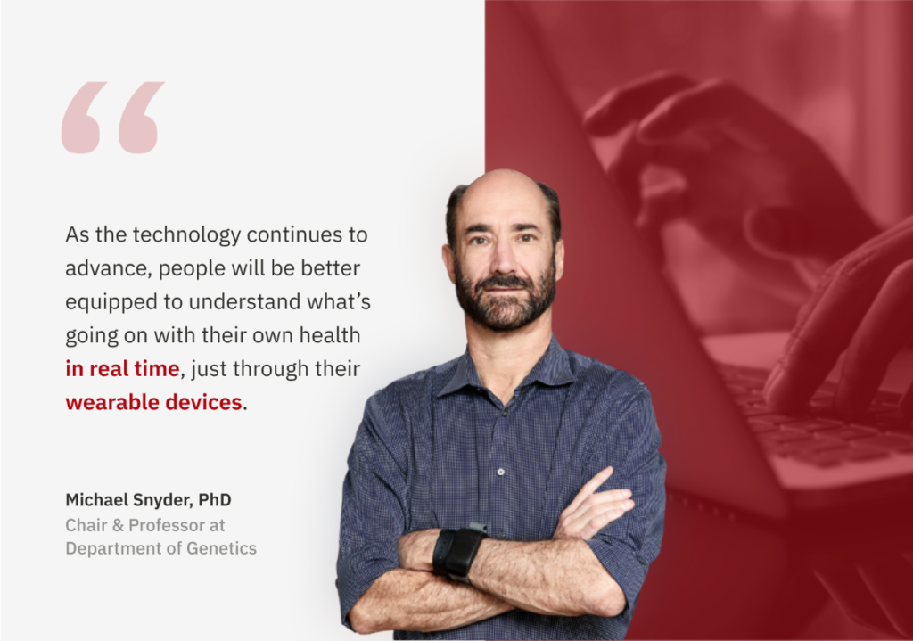
"Stanford School of Medicine professor Michael Snyder is conducting several studies to see how far wearables can go in detecting disease. "You don't drive your car around without a dashboard," he said. "Yet, here we are as people. We're more important than cars, but we're running around without any sensors, most people. And we should be wearing these things, in my opinion, because they can alert you to early things."

Media Spotlight News
Illustration of Michael P. Snyder from Body Count Article in Stanford Magazine
Body Count, how Michael Snyder’s self-monitoring project could transform human health

Body Count, how Michael Snyder’s self-monitoring project could transform human health

"You prick your fingertip, put it on a sensor, and more biometrics scroll along the glass, all in blue, none in red to flag an anomaly."

"This is the future that Michael Snyder, director of the Center for Genomics and Personalized Medicine and chair of the department of genetics, wants for everyone on Earth. He’s already cobbling it together for himself. Each morning, he straps on four smartwatches and an Exposometer to measure levels of airborne particles. He has a continuous glucose monitor for his blood sugar and an Oura ring to track his sleep." Read More >

New Publication
New machine learning algorithm from Stanford team leads to the discovery of hundreds of potential drug targets for ALS

New machine learning algorithm from Stanford team leads to the discovery of hundreds of potential drug targets for ALS

Utilizing a new machine learning method named RefMap, Dr. Michael Snyder and scientists at Stanford Medicine identified hundreds of ALS risk genes, leading to potential new ALS treatment discovery and a 5-fold increase in recovered heritability, a hallmark pathology of ALS.

COVID-19
Graphic showing a smartphone in someone's hand opened to MyPHD study app that's collecting data from a smartwatch
Stanford’s large-scale, real-time monitoring & alerting system detects 80% of COVID-19 illnesses at or prior to symptom onset

Stanford’s large-scale, real-time monitoring & alerting system detects 80% of COVID-19 illnesses at or prior to symptom onset

Harnessing wearable data, the first large-scale, real-time monitoring & alerting system from Stanford detects abnormal physiological events, such as COVID-19 and other respiratory illnesses.

New Publication
Longitudinal data study shows gut microbiome diversity changes at the subspecies level during antibiotic treatment

Longitudinal data study shows gut microbiome diversity changes at the subspecies level during antibiotic treatment

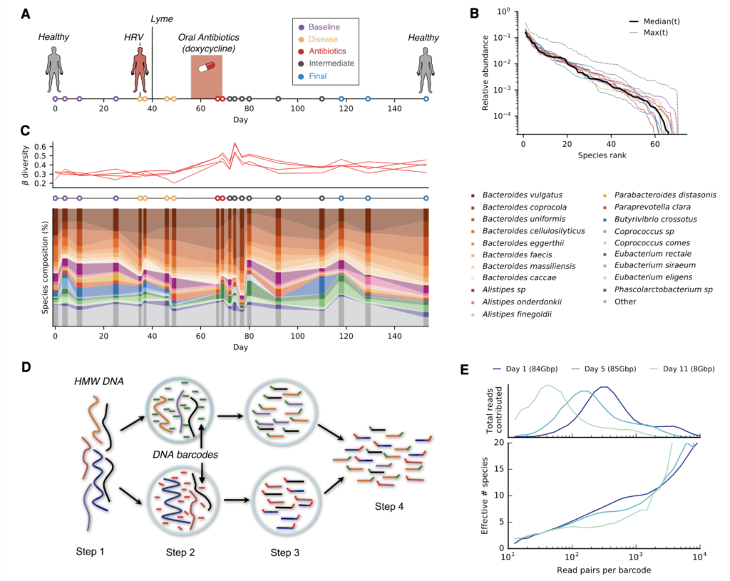
Published as the August 2021 Genome Research cover publication, a Stanford research team found evolutionary & ecological changes in human gut microbiome subspecies during antibiotic treatment based on longitudinal data.

Media Spotlight
Dr. Michael Snyder discusses deep profiling for personalized medicine on Rhonda Patrick’s FoundMyFitness Podcast

Dr. Michael Snyder discusses deep profiling for personalized medicine on Rhonda Patrick’s FoundMyFitness Podcast

Dr. Michael Snyder, a pioneer in longitudinal profiling and personalized medicine, speaks with Rhonda Patrick about his team's work at Stanford, covering areas such as CGMs, multiomics, and wearable data.

Program Launch
A virtual workshop experience from Stanford Medicine featuring topics such as personalized medicine, big data, & AI
Personalized Medicine, Big Data, & AI Explorers, a new virtual summer workshop from Stanford Medicine

Personalized Medicine, Big Data, & AI Explorers, a new virtual summer workshop from Stanford Medicine

A virtual, two-week summer intensive workshop from Stanford Medicine for students 16 and older that transports participants to the forefront of healthcare innovation research featuring topics such as precision health, big data, & AI.

New Publication
Graphic showing a smartphone in someone's hand opened to MyPHD study app that's collecting data from a smartwatch
Real-time alerting system for COVID-19 using wearable data – Stanford preprint available now

Real-time alerting system for COVID-19 using wearable data – Stanford preprint available now

Our preprint for Phase 2 of our COVID-19 wearable study is available now! This is the 1st large-scale real-time monitoring and alerting system for detecting abnormal physiological changes, including COVID-19 and other respiratory illnesses, using smartwatch data.

Research Study
Clinician giving a vaccination
A new research study from Stanford Medicine featuring molecular profiling of COVID-19 vaccine response utilizing at-home microsampling

A new research study from Stanford Medicine featuring molecular profiling of COVID-19 vaccine response utilizing at-home microsampling

A new study from Stanford Medicine aims to understand the differences in individual responses to COVID-19 infection and response to vaccination using microsampling.

Community Events
cloud computing
Insights in the future of cloud computing and its impact on healthcare applications

Insights in the future of cloud computing and its impact on healthcare applications

Dr. Amir Bahmani, Director of Science & Technology at our lab, discussed the future of cloud computing and its impact on healthcare applications with Eric Schmidt, co-founder of Schmidt Futures and former CEO of Google, and Mark Russinovich, CTO of Microsoft Azure.

Become a lab insider.

Sign up for our newsletter and be the first
to hear about new studies in development.

subscribe

Subscriber Benefits

Home

About

Events

COVID-19 Wearables

SCGPM

Stanford Medicine

Healthcare Innovation Lab at Stanford University School of Medicine.
© Stanford University. Stanford, California 94305.